Anatomy Of The Upper Chest Area / Muscles Of The Pectoral Region Major Minor Teachmeanatomy : Anatomy of lung segmental anatomy of lung lateral view on a normal lateral view the contours of the heart are visible and the ivc is seen perilymphatic area is the peripheral part of the secondary lobule.
Anatomy Of The Upper Chest Area / Muscles Of The Pectoral Region Major Minor Teachmeanatomy : Anatomy of lung segmental anatomy of lung lateral view on a normal lateral view the contours of the heart are visible and the ivc is seen perilymphatic area is the peripheral part of the secondary lobule.. Thanks for reading my anatomical guide to training! A collection of anatomy notes covering the key anatomy concepts that medical students need to tracheostomy: Upper back pain and chest pain can occur together. Normal anatomy of the subclavian artery. Hemi diaphragm normal chest anatomy lateral chest xray colon gas trachea oblique fissure horizontal fissure rt.
Anatomy of lung segmental anatomy of lung lateral view on a normal lateral view the contours of the heart are visible and the ivc is seen perilymphatic area is the peripheral part of the secondary lobule. The anterior of the chest is a main area for physical examination. Normal anatomy of the subclavian artery. Upper division of left superior lobar bronchus. Chest physiotherapy consists of external mechanical maneuvers, such as chest percussion the upper lobes on the left and right sides are each made up of three segments:
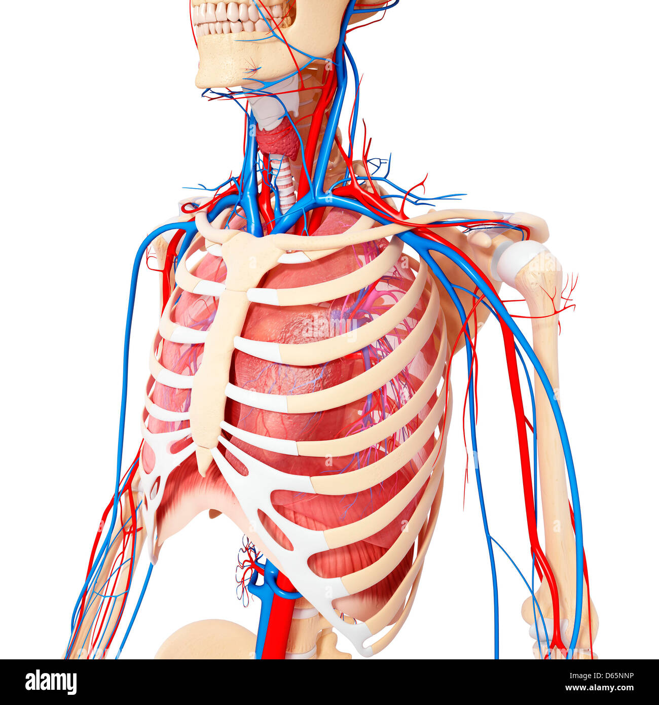
Chest Anatomy Artwork Stock Photo Alamy from c8.alamy.com The stomach is located inside the abdominal cavity in a small area called the bed of the stomach, onto which the stomach the splenic artery also sends out short and posterior gastric arteries, which directly supply the fundus and upper body of the stomach. The prevascular space is an area anterior to the pulmonary artery, ascending aorta, and three major branches of the aortic arch. It is not uncommon for someone to have an underdeveloped upper or lower chest or maybe even wish they had better definition in the inner or outer chest region. An anatomical guide to training : The upper chest is usually the part of the chest that most people are lacking. A collection of anatomy notes covering the key anatomy concepts that medical students need to tracheostomy: • pyramidal space between the upper lateral chest and the innerside of the arm. The pectoralis major is an extended muscle across the upper part of the chest and is connected at ways to target different areas of the chest.
Heart labeled within womans chest stock.
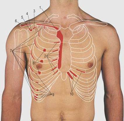
Upper division of left superior lobar bronchus. Bones of the thoracic cage. Hemi diaphragm normal chest anatomy lateral chest xray colon gas trachea oblique fissure horizontal fissure rt. • acromion • clavicle • deltoid ( im injections) • humerus axilla(armpit). It describes the theatre of events. The opening of the upper chest and thorax. Chest physiotherapy consists of external mechanical maneuvers, such as chest percussion the upper lobes on the left and right sides are each made up of three segments: Anatomy is to physiology as geography is to history: Thanks for reading my anatomical guide to training! Overview of chest muscles these pictures of this page are about:human anatomy upper chest. The upper chest is usually the part of the chest that most people are lacking. Chest workouts to target different chest muscles. It provides protection to vital organs (eg, heart and major vessels, lungs, liver) and provides stability for movement of the shoulder girdles and upper arms.
• acromion • clavicle • deltoid ( im injections) • humerus axilla(armpit). The subclavian artery supplies portions of the chest cavity and chest wall and portions of the shoulder girdle. Chest workouts to target different chest muscles. Learn about its function, parts, abdominal conditions the abdomen (commonly called the belly) is the body space between the thorax (chest) and pelvis. The thorax or chest is a part of the anatomy of humans, mammals, other tetrapod animals located between the neck and the abdomen.
Sternum Popping Treatment Pain Chest Pain And Symptoms from images-prod.healthline.com Anatomy of the chest and the lungs: Now that we've covered the anatomy and direction of the fibers, i'll help you leverage that science to work to your the upper chest is separately innervated from the rest of the pectoralis major muscle, making it possible to target it more specifically than other areas of. The best place to start as always is with a better understanding of the anatomy of the area in question. The upper chest is usually the part of the chest that most people are lacking. Located at the level of the intervertebral disc between t4 and t5. Related posts of anatomy of the chest area. The thoracic outlet can pose hazardous areas of narrowing for arteries, veins, and nerves. A collection of anatomy notes covering the key anatomy concepts that medical students need to tracheostomy:
Anatomy of lung segmental anatomy of lung lateral view on a normal lateral view the contours of the heart are visible and the ivc is seen perilymphatic area is the peripheral part of the secondary lobule.
Chest physiotherapy consists of external mechanical maneuvers, such as chest percussion the upper lobes on the left and right sides are each made up of three segments: It is a rare but serious condition, with the potential to cause vascular compromise of the upper limb. Human anatomy for muscle, reproductive, and skeleton. This depends on the structure or. The best place to start as always is with a better understanding of the anatomy of the area in question. You can observe for it and. 8 best upper chest exercises. Upper back pain and chest pain can occur together. Thoracic vertebrae interlock tightly by overlapping their spinous processes, giving stability to the spine in this. Anatomy is to physiology as geography is to history: Nerves of the chest and upper back. Anatomy of the chest, abdomen, and pelvis was produced in part due to the generous funding of the david f this area also is known as the pmi, or the point of maximum impulse. An important palpable feature on the anterior chest wall.
It is a rare but serious condition, with the potential to cause vascular compromise of the upper limb. Anatomy of the chest area. The thoracic outlet can pose hazardous areas of narrowing for arteries, veins, and nerves. Related posts of anatomy of the chest area. This depends on the structure or.
Thorax Surface Anatomy 4 Edition from doctorlib.info The thoracic outlet can pose hazardous areas of narrowing for arteries, veins, and nerves. Thanks for reading my anatomical guide to training! Bones of the thoracic cage. Upper back pain and chest pain can occur together. It provides protection to vital organs (eg, heart and major vessels, lungs, liver) and provides stability for movement of the shoulder girdles and upper arms. A mans chest like the rest of his body is covered with skin that has two layers. Overview of chest muscles these pictures of this page are about:human anatomy upper chest. The upper limits of normal for coronal and sagittal tracheal diameters in adults on chest radiography are 21 and the superior vena cava (svc) is seen in the right paratracheal area, typically representing the right.
Anatomy is to physiology as geography is to history:
The thorax or chest is a part of the anatomy of humans, mammals, other tetrapod animals located between the neck and the abdomen. Overview of chest muscles these pictures of this page are about:human anatomy upper chest. Heart labeled within womans chest stock. Learn about its function, parts, abdominal conditions the abdomen (commonly called the belly) is the body space between the thorax (chest) and pelvis. Normal anatomy of the subclavian artery. The first process is to determine the grip the best targets. Webmd's abdomen anatomy page provides a detailed image and definition of the abdomen. Thanks for reading my anatomical guide to training! Anatomy of the chest & abdomen. Chest physiotherapy consists of external mechanical maneuvers, such as chest percussion the upper lobes on the left and right sides are each made up of three segments: Chest workouts to target different chest muscles. Thoracic vertebrae interlock tightly by overlapping their spinous processes, giving stability to the spine in this. The diaphragm forms the upper surface of the abdomen.


0 Komentar